
The knee is the largest joint of the body and is stabilized by a set of ligaments. In the knee there are four primary ligaments anterior cruciate ligament (ACL), posterior cruciate ligament (PCL), medial collateral ligament (MCL) and lateral collateral ligament (LCL). More complex interactions between a number of other ligamentous/tendinous structures form the posteromedial and posterolateral corners (PMC/PLC)
The lateral collateral ligament (LCL) may tear due to trauma, sports injuries, or direct blows to the knee. Rupture of the LCL may result in pain, swelling and even instability of the knee. LCL injuries can be diagnosed with thorough physical examination and by employing imaging techniques such as X-ray and MRI scan.
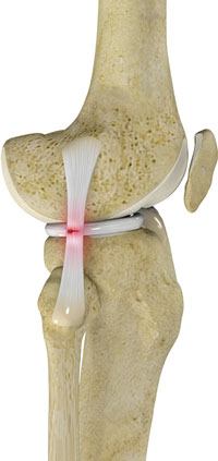
The lateral collateral ligament (LCL) is an important stabiliser present on the outer side of the knee, connecting the thighbone (femur) to the fibula (side bone of lower leg). It provides stability as well as limiting sideways movement of the knee. Tear or injury of the LCL may cause instability of the knee that can be either reconstructed or repaired to regain the stability of the knee.
The initial treatment of the torn LCL include non-surgical interventions such as rest, ice, elevation, bracing and physical therapy to help reduce swelling, and regain activity as well as strength and flexibility of the knee. Surgery is recommended if non-surgical interventions fail to provide stability or if the initial injury is significant. Surgical interventions include repair and reconstruction of the torn ligament. Based on the severity and location of the injury, repair or reconstruction of the LCL is recommended. If the ligament is torn in the middle or if the injury is older than 3 weeks, LCL reconstruction is recommended.
Procedure
An LCL reconstruction involves the replacement of the torn ligament with healthy strong tissue or graft. The tissue or graft can be taken either from the tissue bank (called allograft) or from the patient’s body (called autograft). The type of graft used, depends upon the condition of the patient and choice of your surgeon. Hamstring tendons are commonly used as autograft. A small incision is made on the lateral side of the knee to perform the LCL reconstruction. The procedure is done through an open incision and not arthroscopically. The thighbone and fibula bones are drilled precisely and accurately with specialized instruments to establish bone tunnels. The ends of the tendon graft are passed through these tunnels and are fixed by using screws, metal staples or large sutures. A knee that has undergone LCL reconstruction surgery is braced for 6-8 weeks. A reconstruction of the posterolateral corner (PLC) will often be required at the same time if injured.
Post-operative care
The common post-operative instructions for LCL reconstruction are:
- Use crutches to avoid full weight on the knee for at least 6 weeks
- Use ice and the prescribed medications to reduce swelling
- Avoid lifting heavy weight or vigorous exercise
- Follow the specific instruction given by your surgeon
- Follow rehabilitation programs or work with a physiotherapist to regain the motion and strength of the knee